
The Challenge of Healing in Complex Biological Systems
Medical technologies continue to evolve toward increasingly sophisticated interactions with the body. Whether the objective is tissue repair, localized therapeutic delivery, improved implant integration, or regenerative healing, material performance is no longer defined solely by chemistry or mechanical strength. Increasingly, success depends on how effectively a material interacts with the biological environment in which it is placed.
The challenge is that biology is architecturally complex.
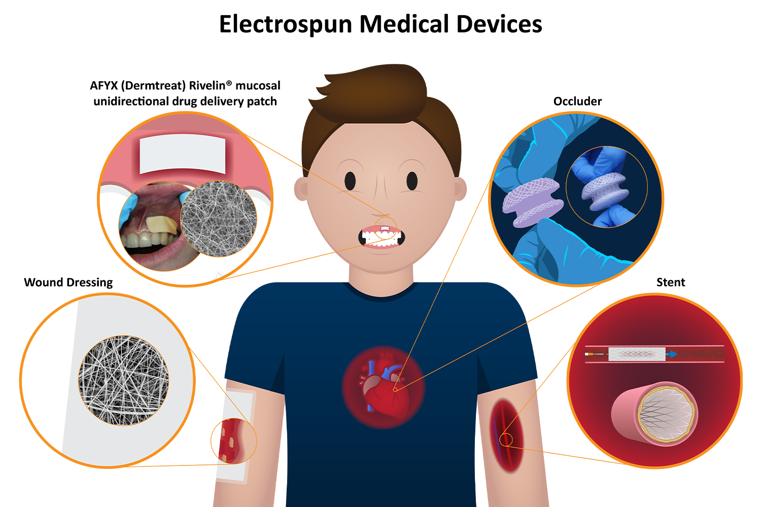
Tissues are not uniform environments. They are layered, porous, mechanically dynamic, and highly specialized to a specific function. Blood vessels balance flexibility with nutrient transport and long-term durability. Peripheral nerves rely on organized pathways to support regeneration. Soft tissue healing requires a coordinated balance between cellular infiltration, permeability, and barrier function. Even implantable devices must integrate into highly variable biological environments while maintaining predictable mechanical performance, some examples of such devices are shown in Figure 1.
Traditional manufacturing approaches have enabled remarkable advances in medical devices and biomaterials, but they often force tradeoffs. Dense polymer systems may provide mechanical durability while limiting cellular interaction and tissue infiltration. Metallic structures deliver strength and dimensional control but offer little biological functionality at the tissue interface. Conventional coatings may modify surfaces yet struggle to conform to complex geometries or meaningfully influence biological response.
As these challenges become more pronounced, materials must evolve beyond static structures toward architectures capable of supporting biological function.
Electrospinning as a Platform for Biological Architecture
Electrospinning offers a fundamentally different approach.
Rather than relying solely on bulk material properties, electrospinning enables materials to be constructed from a tunable fibrous infrastructure that supports architectures designed around biological function. Fiber diameter, morphology, orientation, porosity, and material thickness can all be engineered to influence permeability, degradation behavior, tissue interaction, and therapeutic performance. These variables directly influence how a material performs in vivo and provide a level of design flexibility that is difficult to achieve through conventional manufacturing approaches.
The ability to couple a tunable fibrous infrastructure with application-specific architectures is what makes electrospinning particularly well suited to complex biological systems.
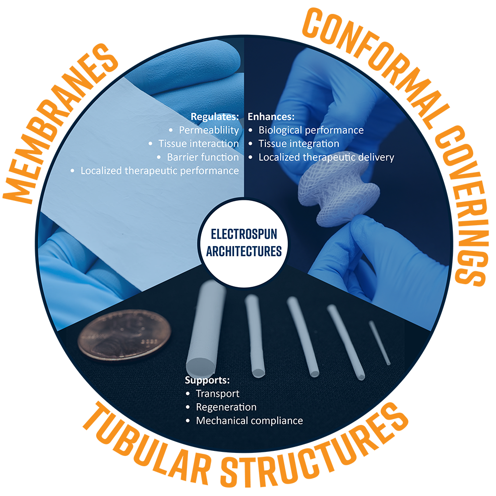
These architectures generally fall into three functional categories:
- Membranes – engineered interfaces designed to regulate permeability, tissue interaction, barrier function, and localized therapeutic performance.
- Tubular Structures – conduit-like geometries intended to support transport, regeneration, and mechanical compliance in systems such as vascular or nerve repair.
- Conformal Coverings – thin functional layers that contour to complex implant geometries to enhance biological performance, tissue integration, or localized therapeutic delivery.
Together, these approaches demonstrate how electrospinning can adapt a common fibrous infrastructure to fundamentally different biological challenges while maintaining control around the intended clinical objective, summarized in Figure 2.
Engineering Controlled Interfaces: Electrospun Membranes
Electrospun membranes remain the most established and widely utilized form factor in electrospinning. Produced as fibrous sheets, membranes have long served as a foundation for applications ranging from wound healing and tissue regeneration to filtration, barrier materials, and localized therapeutic delivery because of their ability to bridge competing requirements at a biological interface.
At their core, electrospun membranes solve an interface problem. In wound healing applications, materials must protect damaged tissue while maintaining oxygen transport and fluid management. Regenerative membranes may need to encourage tissue integration while controlling permeability and degradation. In each case, the challenge lies in balancing competing biological requirements at the material interface.
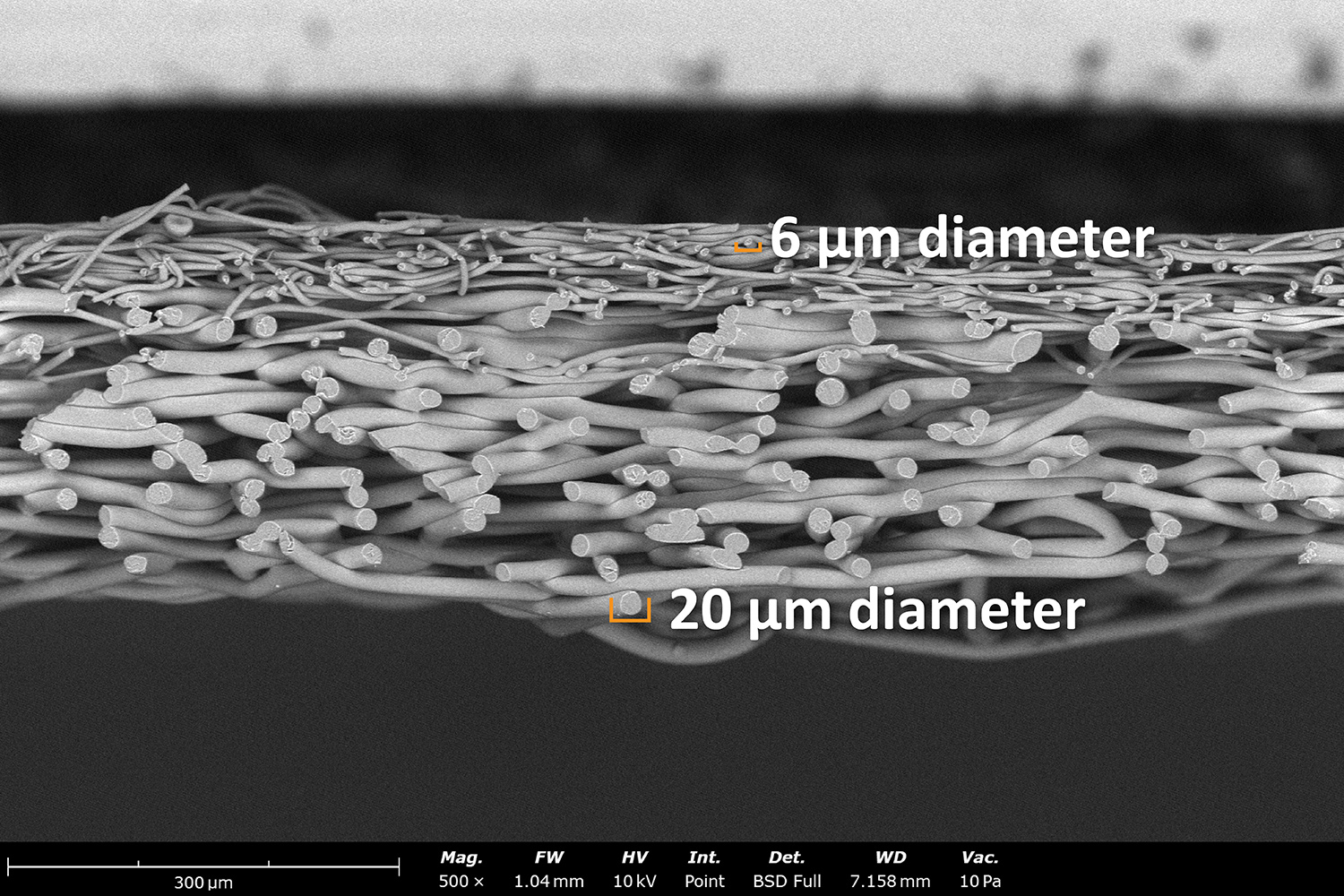
Electrospinning enables these interfaces to be designed intentionally. Fiber morphology, porosity, material selection, and deposition architecture can all be tuned around the intended biological objective. An electrospun bi-layer membrane demonstrating the precision of this technique is visible in Figure 3. Membranes can be engineered for permeability, barrier function, tissue interaction, or localized therapeutic performance depending on the application. Material thickness can also be controlled through deposition time, introducing a meaningful third dimension to scaffold design. Thin films on the order of ~50 µm may be leveraged where flexibility, permeability, or conformability are critical, while thicker architectures in the 2 – 3 mm range can provide greater fluid management, structural integrity, or regenerative support.
From a manufacturing perspective, membrane architectures represent one of the most mature electrospinning pathways. Flat plate collectors and single-needle systems can rapidly generate membrane materials, while rotating drum collectors improve deposition consistency and uniformity across larger areas, commonly producing membranes on the order of ~30 cm × 60 cm. As throughput demands increase, roll-to-roll electrospinning platforms enable continuous membrane manufacturing at widths of approximately 50 cm, producing materials as continuous rolls with tunable thicknesses and lengths. At commercial scales, systems can expand to widths approaching 1.6 meters, enabling large-scale production while maintaining control over the underlying membrane architecture.
This combination of tunable biological performance, controllable geometry, and scalable manufacturing is one of the reasons electrospun membranes continue to serve as a foundational architecture for wound healing, regenerative medicine, and advanced therapeutic materials.
Mimicking Native Biological Conduits: Tubular Electrospun Structures
Electrospun tubular structures represent one of the earliest translational applications of electrospinning, originally gaining attention as architectures for small-diameter arterial grafts where traditional materials often struggled with long-term patency and biological integration. By depositing fibers onto rotating mandrels, electrospinning enables seamless tubular geometries designed to mimic native conduit-like tissues for applications ranging from vascular grafts and nerve repair to soft tissue regeneration and localized therapeutic delivery. Examples of various sizes of electrospun tubes can be seen in Figure 4.
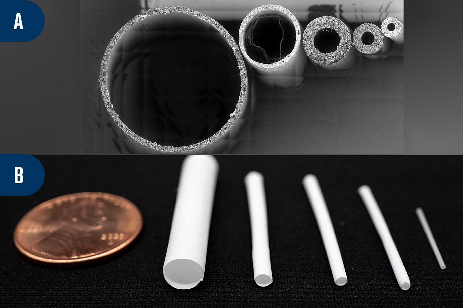
Many biological systems rely on conduits to guide transport, regeneration, or mechanical function. Blood vessels must maintain flexibility under cyclic loading while supporting nutrient exchange, while peripheral nerves require structural guidance to support regeneration across damaged regions. In each case, performance depends not only on geometry, but on how the surrounding material interacts with tissue throughout healing.
Electrospinning enables these variables to be engineered simultaneously. Inner diameter can be tailored to the intended anatomy, ranging from small conduits for nerve repair or microvascular applications to larger diameter structures for vascular interfaces. For vascular applications in particular, electrospun architectures can be designed to balance flexibility and mechanical integrity in ways that reduce the likelihood of kinking while maintaining conduit patency. At the same time, porosity and morphology may be engineered to support tissue integration and healing at the material interface.
Wall thickness can also be tuned to support specific functional requirements, influencing flexibility, suture retention, permeability, and mechanical durability. Thin-walled architectures may prioritize flexibility and transport, while thicker structures can provide greater mechanical support or regenerative function. When paired with tunable fiber morphology and material selection, wall thickness becomes an additional design parameter for tailoring overall material performance.
Tubular architectures are also highly adaptable in scale and length. Short conduit sections may be produced for localized regenerative applications, while vascular grafts can extend to lengths of ~40 cm depending on the intended anatomy. Needle translation along the mandrel length enables more uniform fiber deposition, improving architectural consistency across longer structures.
From a manufacturing perspective, tubular electrospinning presents a different scaling challenge than planar membranes. Because tubules are typically produced on individual rotating mandrels, manufacturing is often performed one conduit at a time rather than through continuous roll-based architectures. However, throughput can still be highly practical depending on the intended geometry and electrospinning configuration. With appropriate needle configurations and optimized deposition conditions, tubular architectures can often be manufactured in minutes rather than hours while maintaining control over wall thickness, morphology, and overall geometry.
This balance between tunable geometry, rapid throughput, and architectural control is one of the reasons electrospun tubules continue to gain traction across vascular repair, nerve regeneration, and regenerative medicine applications.
Enhancing Existing Platforms: Electrospun Device Coverings
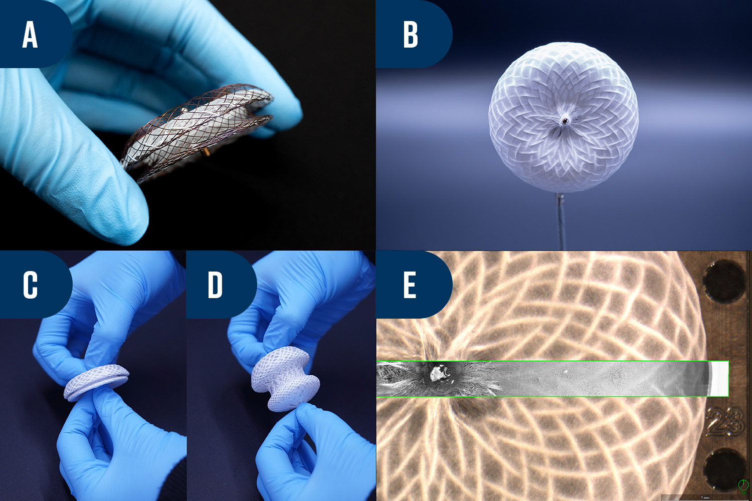
Electrospun coverings extend the architectural flexibility of electrospinning directly onto existing medical devices. By depositing fibers conformally onto complex geometries, electrospinning enables thin, porous layers to contour to open structures, irregular surfaces, and catheter-deliverable implants while introducing biological functionality at the material interface.
This approach is particularly well suited to metallic implant frameworks such as nitinol laser-cut devices used in stents, occluders, vascular grafts, and structural heart applications. In these systems, the metal frame provides structural support and deployment mechanics, while the electrospun layer engineers the biological interface.
Device coverings themselves are not new. Expanded PTFE, woven fabrics, and polymer films have long been used to introduce barrier functions or improve implant performance. However, these approaches often require multiple manufacturing and assembly steps including wrapping, suturing, lamination, or secondary attachment processes. Electrospinning enables functional layers to be deposited directly onto complex geometries in a single step while simultaneously introducing tunable porosity, therapeutic loading, and biological functionality.
Electrospun coverings can be tailored around competing performance requirements. Thin conformal layers may prioritize flexibility and low-profile delivery for minimally invasive systems, while thicker architectures can support greater tissue interaction, barrier function, or therapeutic loading. When paired with tunable fiber morphology and material selection, coverings can be engineered to balance permeability, durability, healing response, and mechanical performance around the intended application.
One of the strongest advantages of electrospun coverings is the ability to move beyond passive coverage. Active pharmaceutical ingredients may be incorporated directly into the fibrous structure to create localized drug-eluting systems, while porosity and scaffold morphology may be engineered to support tissue integration and controlled healing. For long-term implants, multilayer architectures, surface modification strategies, and post-processing approaches can further improve integration between the electrospun layer and the underlying device.
This combination of structural compatibility, biological functionality, and single-step deposition onto complex geometries is one of the reasons electrospun coverings continue to gain traction across cardiovascular devices, regenerative implants, and next-generation therapeutic platforms such as the occluder device shown in Figure 5.
Enabling Architectures for Healing
The promise of electrospinning is not simply the ability to manufacture fibrous materials, it is the ability to design material architectures around a biological objective. Whether the challenge is balancing permeability and tissue integration in a membrane, supporting regeneration through a conduit, or enhancing the biological performance of an implant through a conformal covering, electrospinning enables material systems to be tailored around how the body heals and responds. The challenge lies in translating these concepts into materials that perform predictably in a real-world application, where material selection, fiber morphology, deposition strategy, and form factor must all align with the intended therapeutic or device objective.
At Nanoscience Analytical, we work alongside customers to develop electrospun materials around specific technical and biological requirements. From early-stage feasibility and material screening to process development and architecture optimization, our team supports programs focused on regenerative medicine, implantable devices, wound care, and advanced therapeutic systems.
Whether you are evaluating an electrospun device covering, regenerative scaffold, or drug-eluting material platform, our team can help identify the right material systems and electrospinning strategies to move your technology forward.